
Scientists from Trinity School Dublin and the Royal School of Surgeons in Eire have developed progressive fluorescent dyes that change colour to visualise totally different organic environments utilizing a single dye. These dyes, able to “switching on” and “off” primarily based on their location inside mobile constructions, allow real-time, high-contrast imaging of mobile processes. This breakthrough, revealed within the journal Chem, paves the way in which for developments in bio-sensing, drug supply imaging, and the research of mobile dynamics. The analysis advantages from worldwide collaboration and vital funding from Irish analysis our bodies, promising a variety of purposes in biology and medication. Credit score: SciTechDaily.com
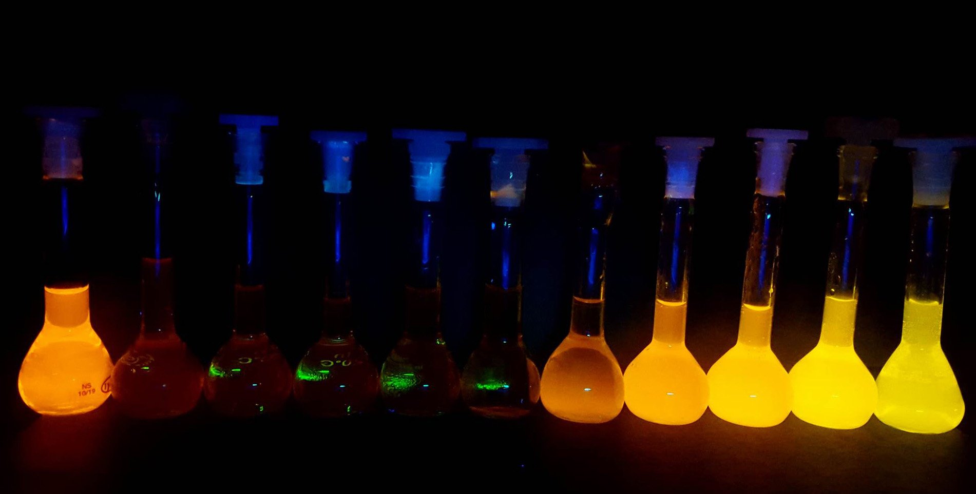
Researchers at Trinity School Dublin, working along with the Royal School of Surgeons in Eire (RCSI), have developed particular fluorescent, color-changing dyes that, for the primary time, can be utilized to concurrently visualize a number of distinct organic environments utilizing just one singular dye.
When these dyes are encapsulated in supply vessels, like these utilized in applied sciences just like the COVID-19 vaccines, they “change on” and provides out mild through a course of referred to as “aggregation-induced emission” (AIE). Quickly after supply into the cells their mild “switches off” earlier than “switching on” once more as soon as the cells shuttle the dyes into mobile lipid droplets.
Superior Imaging Methods
As a result of the sunshine coming from contained in the cells is of a unique colour and happens inside a unique time window to the sunshine coming from the identical dye contained in the supply vessels, the researchers can use a way referred to as “fluorescence lifetime imaging” (FLIM) to differentiate between the 2 environments in real-time.
The work was not too long ago revealed within the main worldwide journal, Chem. First writer, Dr Adam Henwood, Senior Analysis Fellow within the Faculty of Chemistry and primarily based on the Trinity Biomedical Sciences Institute (TBSI), labored on this design with PhD pupil Connie Sigurvinsson.
Dr Henwood defined: “Bioimaging depends on “on/off” dyes the place the dyes solely emit mild underneath one set of situations however are in any other case switched off. That is extraordinarily helpful, nevertheless it does imply you can solely have a look at one place at a time underneath your microscope. The thrilling half about this work is that our dyes hit a candy spot that provides them distinctive on/off/on properties and, crucially, we are able to each observe and differentiate these totally different “on” states.
“So, we each see extra and see higher than earlier than. We do that by timing how lengthy it takes for the sunshine coming from our samples to succeed in the microscope: mild from the supply vessels takes marginally extra time than mild from throughout the cells. By accumulating sufficient mild alerts, we are able to use this info to quickly construct up exact 3D photographs of the 2 totally different dye environments. The time variations are small – only a few billionths of a second both approach – however our technique is delicate sufficient to seize it.”
This distinctive high quality means the dyes may have an enormous suite of purposes and, for instance, maintain the potential to revolutionize bio-sensing and imaging approaches.
Luminescence adjustments of the identical dye shifting from pure natural solvent, left, to water, proper. Credit score: Dr Adam Henwood, Trinity School Dublin
As a result of these dyes may help scientists map the intricate constructions inside residing cells with such excessive distinction and specificity, they might assist illuminate how medication are taken up and metabolized by cells or enable scientists to design and conduct a variety of latest experiments to higher our understanding of the advanced internal workings of cells and their all-important biochemical equipment.
Within the revealed journal article, the scientists centered on utilizing the dyes to picture mobile lipid (fats) droplets, that are one instance of essential “organelles” that make up residing cells in most advanced organisms (like us people).
Lipid droplets, as soon as thought of to be easy “fats reservoirs”, are actually believed to play an essential function in regulating mobile metabolism, coordinating lipid uptake, distribution, storage, and use within the cells. Due to this rising understanding of their significance, and since sudden adjustments of their exercise usually point out mobile stress, they function a helpful check case situation for the dyes. One potential avenue of additional analysis is to see whether or not the group can goal different essential mobile organelles with their dyes.
Thorfinnur Gunnlaugsson, Professor of Chemistry within the Faculty of Chemistry at Trinity and primarily based in TBSI, is the senior writer of the article. He mentioned:
“Having the ability to monitor mobile operate or the movement of molecules or drug candidates inside cells by observing totally different fluorescence emission colours is extraordinarily engaging. The breakthrough right here is that we are able to resolve and use the distinction of their fluorescence lifetimes to establish these similar probes inside totally different mobile environments in a quick and correct method, actually permitting us to map out their colourful “time journey” throughout the cells.
“Most enjoyable, nonetheless, is that this phenomenon doesn’t apply to mobile imaging. These outcomes open up new potentialities in all the pieces from learning chemical biology, as we now have proven right here, to many different medical purposes and even within the era of novel useful supplies to be used past biology. Any molecular or nanomaterial that requires managed molecular movement can in precept be mapped and fine-tuned utilizing our new technique.”
Potential Functions and Future Instructions
And certainly, it’s right here the place the authors intend to forged the online far and broad. They envisage many new potentialities for these dyes, pointing towards their distinctive sensitivity as engaging for growing sensors of hazardous environmental pollution or utilizing their vivid, light-emitting properties to energy chemical transformations, analogous to nature’s personal photosynthesis.
The analysis has each a world (eight nations are represented) and Irish really feel to it, with the latter’s key funding our bodies the Irish Analysis Council (IRC) and Science Basis Eire each taking part in important monetary help roles. Most notable is SFI’s Analysis Centre for Prescribed drugs, SSPC, which principally funded the work, in addition to contributions from the SFI AMBER middle and thru the AMBER-based EPSRC-SFI Centre for Doctoral Coaching Programme.
Prof. Damien Thompson, Professor of Physics on the College of Limerick and Director of the SSPCsaid: “As a middle, we preserve pushing ahead and creating new data on the interface of supplies and biology. This collaborative work between two of our principal investigators at Trinity and RCSI showcases the facility of elementary science to drive innovation in medication. The nearer we have a look at the molecule-cell interface, and crucially, the higher we are able to see, in real-time, how molecules diffuse from place to put contained in the cell nanomachinery, the nearer we get to realising Richard Feynman’s dream of understanding all the pieces that residing issues do from the wiggling and jiggling of atoms.
“However solely not too long ago have researchers had enough experimental and computational assets to trace these motions and vibrations in advanced organic environments. This thrilling new work demonstrates extra particular, excessive distinction imaging of subcellular dynamics, which is able to in flip allow researchers to develop simpler drug formulations with diminished negative effects.”
Professor Donal O’Shea, who oversaw the investigation, is an knowledgeable in cell imaging primarily based in RCSI’s Division of Chemistry and Tremendous-Decision Imaging Consortium (funded by Science Basis Eire, SFI). He added: “Our use of FLIM to trace dynamic AIE interactions with residing cells is an strategy that may have broad applicability for different fluorophore techniques permitting insights to be gained that have been beforehand hidden.”
Reference: “Time-resolved fluorescence imaging with color-changing, “turn-on/turn-on” AIE nanoparticles” by Adam F. Henwood, Niamh Curtin, Sandra Estalayo-Adrián, Aramballi J. Savyasachi, Tómas A. Gudmundsson, June I. Lovitt, L. Constance Sigurvinsson, Hannah L. Dalton, Chris S. Hawes, Denis Jacquemin, Donal F. O’Shea and Thorfinnur Gunnlaugsson, 1 December 2023, Chem.
DOI: 10.1016/j.chempr.2023.10.001
The research was funded by the Irish Analysis Council and the Science Basis Eire.
Discover more from PressNewsAgency
Subscribe to get the latest posts sent to your email.